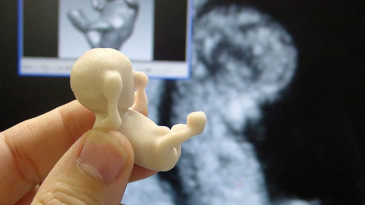
A sonogram (obstetric ultrasonography) allows pregnancy to become more real for the expectant parents. Its intended purpose is to inform patients and doctors about the health of the baby and mother at the beginning of prenatal care. The sonogram is also known to many people as an ultrasound, and its historical moment began in 1963. It advanced 24 years later in 1987 when the very first 3D sonogram was crafted.
Normally in this procedure, sound waves are carried in 2D scanning, sending sounds down and back. However, 3D scanning provides sounds from all angles which are then processed by a computer program, which then reconstructs the information into a 3D image of the fetus and its organs. This provides a more human-like result than the original 2D ultrasound.
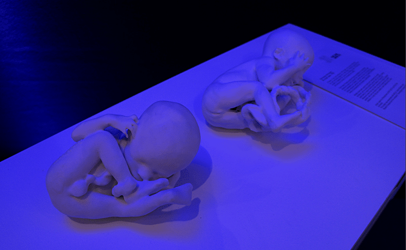
In recent years, there have been incredible advancements to sonograms. It’s flashed forward from black and white specs to a 3D image to now allowing parents to hold a 3D life-size model of their baby while he or she is still in utero. Although sonograms are still used as a screening method to monitor the growth and health of child and mother, nine months is a long time to wait to see and hold the precious child one is carrying.
Therefore, this procedure allows a 3D printer to retrieve the 3D sonogram data and recreate an exact replica of the fetus. It prints 3D sonogram models that can be held by the mom and dad in waiting. This is not only helpful for the parents, but also for doctors too since this can help identify and prepare parents for any deformities as well as provide a visual 3D sonogram for the visually impaired to feel the process of their baby’s growth.
Now you no longer have to wait the entire nine months to see him or her in 3D with this sonogram technology. The Tecnologia Humana 3D will change lives in many ways and will only continue to advance as technology does. I am totally going to go cheesy and say…baby…we have come a long way!
Hold A Life-Size Model Of Your Unborn Baby With This 3D Sonogram
Via: [feto3d]


COMMENTS