While most patients are familiar with the basic tools of radiology like X-rays, CT scans, and MRIs, the field is undergoing exciting new developments each year. These significant advancements show the way toward helping more patients with non-invasive scans of their vital organ systems.
Jose Pizarro, a neuroradiologist based out of Longboat Key, Florida, shares seven recent advancements in the field of radiology and details on how they can help patients with debilitating disorders live more normal lives.
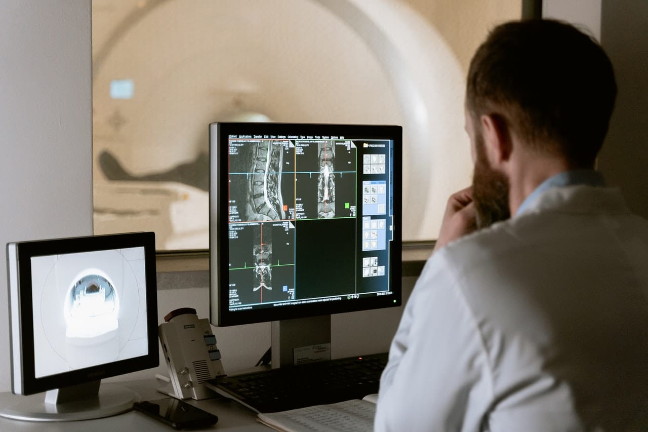
IMAGE: PEXELS
1. Diffusion Tensor Imaging And Fiber Tractography
Diffusion tensor imaging and fiber tractography are used to map the structure of the living human brain. By measuring how water is diffused through the varying types of tissues in the brain, this radiological technique can produce a working model of the structures of white and gray matter.
Diffusion tensor imaging and fiber tractography can help surgeons plan the safest route to reach a brain tumor or other type of lesion. This technique is relatively new and has received many updates in recent years.
2. Angio-CT Technology
The hybrid use of CT scans and fluoroscopy is not often used in the United States, but the technique has been used for some time in Japan. American doctors are beginning to use CT and fluoroscopy combinations together because they can provide greater efficiency and a better patient experience.
One of the best uses for hybrid angio-CT imaging is in interventional oncology. Hybrid imaging allows the doctor to see exactly where they are treating and provides the best depiction of the amount of radiation delivered to keep doses appropriate for the patient.
3. AI And Neuroimaging
AI is becoming a vital part of the medical field. The technology has an incredible potential for adoption in radiology. The future of neuroimaging is carried by AI technologies, which can help doctors come to conclusions faster by quickly analyzing large numbers of images and returning usable data.
AI can be used for triage that can detect a brain hemorrhage before it can normally be detected. It can also help detect strokes, alerting the stroke team automatically when large vessels become blocked.
4. Integrated 3-D Imaging
3-D imaging has a special place in cardiac imaging. 3-D echocardiography has been able to guide doctors in printing models of the heart to inform their interventional plans. Three-dimensional modeling can improve surgical outcomes while decreasing exposure to radiation and making procedures more efficient.
Using only one or two methods of integrated imaging on their own may not be sufficient to truly understand the issues in a patient’s heart. Combining CT, MRI, and 3-D echocardiography can help doctors better understand heart disease.
5. Ultrasound
Ultrasound technology has developed a great deal over the past several years. From 3-D ultrasounds for pregnant women to heart and abdominal imaging, ultrasound is one of the safest types of scan and the most effective for many purposes.
Ultrasound images have improved in clarity thanks to real-time computer imaging. With better images, doctors can improve their rate of accuracy in making a diagnosis.
Volumetric ultrasounds can create images with more detail and depth than traditional ultrasound. These scans can examine cardiac function, identify tumors, and assist in the diagnosis of many conditions. Images produced by volumetric ultrasound are 3-D in nature and can show more detail.
6. Remote Viewing Of Images
Remote viewing of radiologic images has been instrumental in getting patients the care they need. Remote care enables patients who live in less-populated areas to receive care from a well-known radiologist in another city. If the patient is found to have a serious problem that cannot be treated on-site, the patient can be transferred to a larger hospital with more advanced technology.
7. Digital Mammography
In the past, mammograms often had high false positive and false negative readings. Digital mammography has helped doctors to better distinguish between normal tissues and potentially cancerous areas. With digital mammography, patients need to undergo fewer biopsies and other invasive treatments.
8. Better Patient Comfort
Mammograms and other types of diagnostic imaging tests are often viewed as painful and uncomfortable. Companies are beginning to respond to these patient complaints and make their imaging hardware less difficult for patients to interact with. Patients will experience less anxiety and stress and will be more likely to return for future testing if their comfort is considered.
Keeping Track Of A Rapidly Changing Field
Radiologists like Jose Pizarro recognize the necessity of staying abreast of the latest research findings and the newest imaging techniques in the field. Forward-looking physicians are excited to utilize new methods of diagnosing and treating their patients. From ultrasound to 3-D hybrid imaging, patients will receive the best possible treatment with lower exposure to radiation and greater comfort.
If you are interested in even more technology-related articles and information from us here at Bit Rebels, then we have a lot to choose from.
COMMENTS