This is the moment when a surgical shortcut becomes a systems design problem. Neuralink’s announcement that it performed its first transdural procedure in May of 2026 with Dr. Andres Lozano at UHN in Toronto is not merely an incremental tweak to technique.
The real significance here is not that surgeons no longer cut the dura. The part that changes how this should be understood is that preserving the dura turns what used to be an open mechanical problem into a combined optics and precision insertion problem, and those two domains scale differently.
Most people assume less invasive always means simpler engineering. That is a misunderstanding. Skipping a durectomy removes a visible surgical step, but it creates new constraints on needle geometry, imaging, and the forces required to penetrate a leathery membrane while avoiding blood vessels beneath it. What actually determines whether this matters is how well those constraints can be satisfied repeatedly, safely, and at volume.
Neuralink says the transdural approach makes surgery safer, faster, and potentially more scalable. The company moved from opening the dura and exposing the cortex roughly to the size of a quarter to inserting electrode threads directly through the dural layer. To achieve that, the team iterated needle geometry, built synthetic dural membranes for benchtop testing, and added two imaging systems to the robot: ICG video angiography and optical coherence tomography, or OCT.
What becomes obvious when you look closer is this is as much a change in risk profile as it is a change in workflow. You trade a durectomy and direct sightlines for a procedure that depends on dye-driven vasculature contrast and laser depth sensing to guide submillimeter threads. Whether that tradeoff favors broad deployment depends on how the engineering constraints scale across patients, operating rooms, and long-term implant outcomes.
What Transdural Surgery Changes
Transdural surgery replaces cutting the dura with a strategy that threads electrodes through the intact membrane, relying on optics and refined needle mechanics to avoid vessels and place electrodes at cortical depth. Preserving the dura reduces exposure, shortens recovery in some cases, and shifts the problem from open sightlines to imaging and insertion precision.
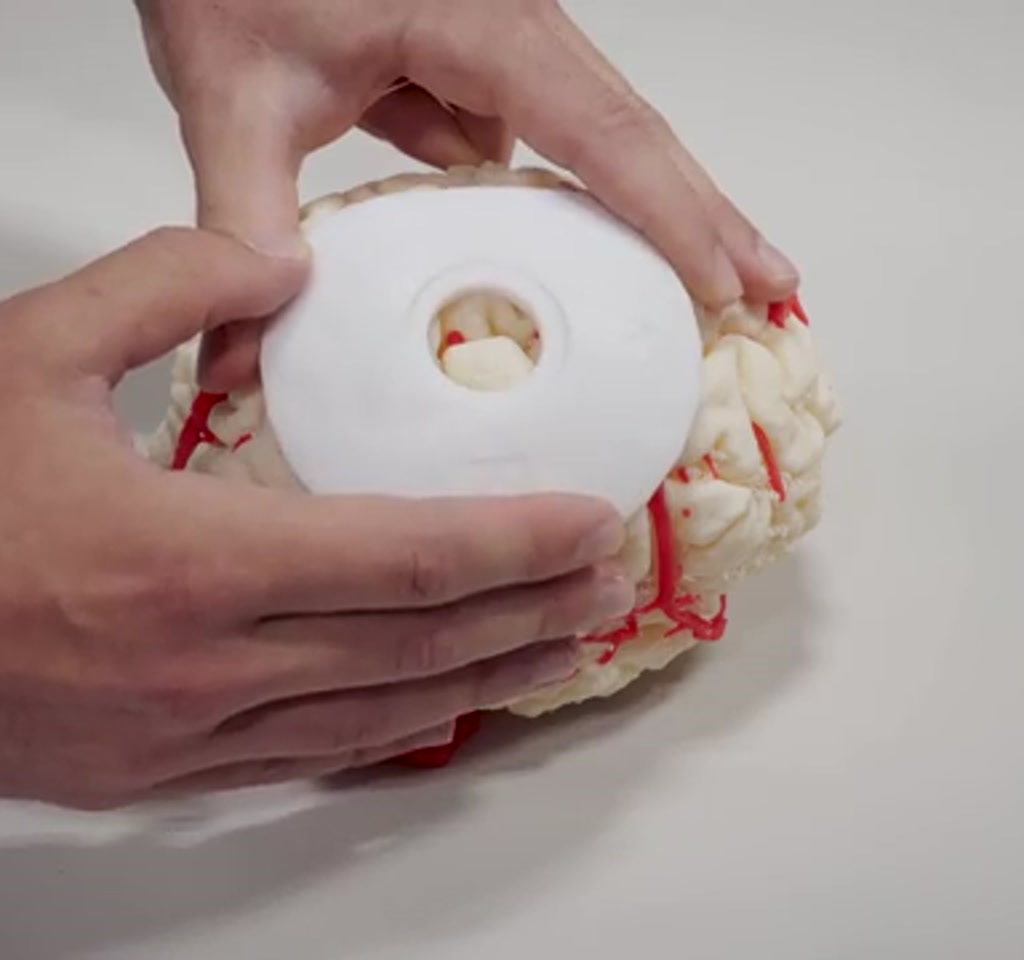
Preserving the dura removes a routine step: opening and excising a patch of the dural membrane so the surgeon can see and work on the cortical surface. Neuralink describes the exposed cortical window in prior procedures as roughly the size of a quarter. With the dura intact, the surgeon and the robot no longer see blood vessels or the exact distance to the cortex by sight alone.
The immediate benefits are tangible. Keeping the dura intact is expected to reduce operative time, lower exposure related infection risk, and shorten recovery for some patients. It also reduces the size of the craniectomy exposure needed for the procedure. That combination makes the operation more compatible with high-throughput, robot-assisted workflows under clinical supervision.
That compatibility is conditional. The approach only holds up when imaging and insertion reliability are high enough to replace direct visual judgment. The threshold for that reliability is set by how accurately the system can map vasculature and cortical depth in real time, and by how consistently the needle and threads traverse the dura without excessive tissue disruption.
Engineering Challenges And Tradeoffs
Transdural implantation reframes constraints into measurable engineering variables: puncture force, needle geometry, vascular mapping fidelity, and depth sensing accuracy. Each variable has tradeoffs that affect immediate safety and long-term electrode performance, and those tradeoffs determine whether the approach scales beyond specialized centers.
Penetrating The Dura
The dura is a tough, leather-like membrane. Neuralink reports their original needle design could not reliably penetrate it. The engineering response was not brute force, but incremental geometry. The needle diameter was increased slightly so electrode threads could be carried through the membrane. That sounds minor, but small changes in diameter on the order of fractions of a millimeter can alter insertion force, tissue stretch, and the profile of the puncture wound.
To measure those effects, the team built an entire testing pipeline. They developed synthetic dural membranes that mimic human thickness and puncture force characteristics, then ran hundreds of insertion tests.
Those hundreds of cycles provided a bounded data set that informed needle shape, bevel angle, and insertion speed. Bench testing at that scale is a practical constraint that many new surgical approaches skip; here it becomes a gating requirement before clinical translation.
Two explicit tradeoffs emerge. Increasing needle diameter improves puncture reliability but raises the chance of acute tissue trauma and chronic scarring along the insertion tract. Second, pushing through the dura rather than cutting it introduces repeated, localized punctures to a membrane that normally remains intact, and the long-term neural tissue response to that pattern of disruption is not yet established.
Seeing Through The Dura
Keeping the dura intact blocks two critical visual cues: blood vessel locations and the distance from the dural surface to the cortical surface. Neuralink solved these with two distinct optical technologies, each carrying its own boundaries.
First, to map vasculature the team used indocyanine green video angiography. ICG is an intravenous dye that fluoresces under infrared illumination, making blood vessels visible even through dura. This creates a temporary, high-contrast view of the vascular map, which the robot uses to steer the threads away from surface vessels.
Second, to measure depth the robot uses OCT. Optical coherence tomography emits low-power infrared light and reconstructs a three-dimensional tomographic image that estimates the distance to the cortex. Neuralink describes feeding that light through fiber optics in the robot head and visualizing a 3D volume of the brain tomographically to control insertion depth in real time.
Both technologies are powerful, but each has limits that become practical constraints. ICG provides a time-limited window of contrast, so angiography must be timed and repeated as needed. While allergic reactions to ICG are rare, the requirement for systemic dye injection introduces an added procedural step and monitoring requirement.
OCT offers high axial resolution, but its penetration depth is typically limited to a few millimeters and the measured distance must be interpreted against brain motion caused by cardiac and respiratory cycles. In practice this means depth estimates vary on the order of millimeters during live surgery, and the system must synchronize insertion timing with that motion or track it continuously.
How A Robotic Workflow Fits In
Robotic workflows aim to trade manual judgement for reproducible instrumentation, making repeated submillimeter insertions feasible under clinician supervision. Deleting the durectomy reduces subjective steps, but only if imaging and insertion repeatability meet clinical thresholds across patient variability and operating environments.
Neuralink frames the transdural step as a way to delete the durectomy from the workflow, making the operation faster and more compatible with robots under clinical supervision. The company envisions robots executing repetitive precision insertions while a clinician supervises higher-level decisions. The deleted step reduces the number of manual surgical actions that require subjective judgement and continuous manual dexterity.
Automation becomes feasible when two conditions are met. First, instrumentation must be robust across patient variability. Neuralink’s hundreds of bench insertions are a start, but clinical diversity in dural thickness, adhesions from prior surgery, and anatomical variance requires broader validation. Second, imaging must reliably resolve vasculature and depth in every case, not just in controlled trials.
The tradeoffs here are operational. If the imaging and insertion systems require specialist operators or frequent calibrations, throughput gains from automation will be limited. Equipment costs and maintenance are also constraints. High-precision OCT modules and fluorescence imaging systems add capital and per-procedure consumable costs, and hospitals will weigh those against the time and safety improvements on a case-by-case basis.
Clinical Trials And The Road Ahead
According to Neuralink, the first transdural procedure under clinical trial conditions took place in May of 2026 at University Health Network in Toronto with Dr. Lozano. That step is a practical milestone which signals transition from benchtop validation to human testing. Clinical trials will need to address patient selection, acute complication rates, and long term outcomes such as electrode stability and tissue response around transdural puncture sites.
There are at least two measurable boundaries to watch during those trials. One is insertion reliability across a patient cohort, which will be evaluated in terms of successful thread placement per attempted insertion and any vascular injury events. The second is the longitudinal performance of implanted electrodes, quantified by signal stability over months to years and by rates of scar formation that could degrade recordings.
Transdural Vs Durectomy: Comparison And Decision Factors
Comparing transdural implantation and traditional durectomy centers decision making on visible exposure, infection risk, procedure time, imaging dependency, and long term tissue response. Transdural reduces open exposure and may cut operating time, while durectomy preserves direct sightlines and avoids repeated punctures of the dura.
Safety And Visibility
Durectomy provides direct visualization of cortical blood vessels and cortical topography, reducing reliance on temporally limited imaging. Transdural procedures substitute that visibility with ICG angiography and OCT, which introduce their own timing and interpretation constraints.
Throughput And Cost
Transdural techniques can shrink per case time but add capital cost for OCT and fluorescence systems plus consumables such as ICG. High volume centers may justify the investment, while lower volume hospitals will balance equipment expense against minutes saved per operation.
Constraints That Define Usefulness
Two concrete constraints shape how far transdural surgery will travel. First, imaging windows and measurement fidelity define a performance envelope. ICG angiography yields a temporary vascular map and OCT provides depth at a few millimeters of penetration.
In aggregate, those systems must keep the robot out of blood vessels while ensuring threads land within a narrow cortical depth window that is often measured in single-digit millimeters.
Second, procedural economics matter. Removing a durectomy reduces a step and likely cuts minutes to tens of minutes from procedure time, which matters for throughput. But the added equipment, consumables such as ICG dye, and maintenance for high-precision optics all introduce costs that tend to scale into the hundreds or thousands per operating room rather than the tens. Hospitals and payers will judge whether the time saved and safety gains justify those ongoing expenses.
Those constraints are not blockers. They are conditions that define where the approach is useful, and they point to the kinds of centers and indications most likely to adopt transdural implantation early: high-volume neurological centers with existing surgical robotics experience and the capital to support advanced imaging suites.
Quotable point: The part that changes how this should be understood is that preserving the dura turns the visual and mechanical problem into an optics problem, and optics scale differently than open surgery.
What becomes clear from Neuralink’s description is that transdural surgery is less a shortcut than a redesign. The company traded a visible surgical window for a layer of engineering that sits between patient and surgeon: synthetic dural proxies, refined needles, ICG-guided vascular maps, and OCT-driven depth control. Each element has its testable limits and its own maintenance and clinical implications.
Two editorial observations follow. One, the approach aligns with a broader trend in neurotechnology where invasiveness is reduced by pushing complexity into instruments and software. Two, that strategy only scales when dynamics such as brain motion, patient variability, and long-term tissue reaction are addressed with data across many patients and time spans that extend beyond initial trials.
The transdural milestone reported in May of 2026 is a technical statement and a roadmap. It indicates which problems were solved and which remain conditional on wider validation. The most interesting question going forward is not whether a robot can thread electrodes through a membrane but whether a health system can reliably, safely, and economically deliver that capability at scale while tracking long-term outcomes.
That is the metric that will determine whether this approach remains a niche within specialized centers or becomes a standard pathway that opens BCIs to many more patients. The next phases of clinical work will reveal the contours of that decision space and the practical thresholds that define usefulness in real-world care settings.
Who This Is For And Who This Is Not For
Who This Is For: High-volume neurological centers with surgical robotics experience, research hospitals conducting clinical trials on BCIs, and teams that can absorb capital costs for OCT and fluorescence imaging. Early adopters will prioritize throughput gains and advanced imaging suites.
Who This Is Not For: Low-volume hospitals without existing robotic surgery programs, centers that cannot support additional imaging consumables, and cases where direct cortical visualization remains essential due to prior surgical scarring or unusual anatomy. Patients and clinicians should await trial results for long-term device performance data before widespread adoption.
Frequently Asked Questions
What Is Transdural Surgery For Brain Implants?
Transdural surgery inserts electrode threads through the intact dural membrane rather than removing a dural patch. It relies on imaging such as ICG angiography for vessel mapping and OCT for depth sensing to guide placement without direct visual access to the cortex.
How Does ICG Angiography Help In Transdural Procedures?
ICG angiography is an intravenous dye technique that fluoresces under infrared light, creating a temporary high-contrast map of surface vasculature that can be seen through the dura. Neuralink uses it to steer threads away from visible vessels during insertion.
What Role Does OCT Play In Needle Insertion?
Optical coherence tomography provides tomographic depth estimates by emitting low-power infrared light and reconstructing a 3D volume. In the transdural workflow OCT estimates distance from the dural surface to the cortex to control insertion depth in real time.
Is Preserving The Dura Safer Than A Durectomy?
Preserving the dura reduces open exposure and may lower infection risk and recovery time, but it introduces different risks related to repeated punctures and imaging dependence. Definitive safety comparisons require the broader clinical trial data that are currently being collected.
Can Robots Perform Transdural Insertions Reliably?
Robotic execution is feasible when imaging and insertion systems are validated across patient variability. Neuralink has completed extensive bench testing and reported an initial clinical procedure, but larger trials are required to confirm reliability across diverse patient populations.
What Are The Main Barriers To Widespread Adoption?
Primary barriers include imaging fidelity limits, long-term tissue responses to repeated dural punctures, equipment and consumable costs, and the need for multi-center trial data showing consistent outcomes across patients and time.
How Will Clinical Trials Measure Success?
Trials will measure insertion reliability, vascular injury rates, electrode signal stability over months to years, and rates of scar formation that could affect recording quality. Patient selection and acute complication monitoring will also be key metrics.
Does The Current Reporting Resolve Long-Term Outcomes?
No. The May 2026 transdural milestone is an important technical and clinical step, but long-term outcomes such as chronic tissue response and sustained signal quality need longitudinal data from expanded clinical trials before conclusions can be drawn.
COMMENTS